


您的位置:首页 > 产品中心 > Anti-Cytochrome P450 26A1 Antibody, clone F27P6A1
Anti-Cytochrome P450 26A1 Antibody, clone F27P6A1

产品别名
Anti-Cytochrome P450 26A1 Antibody, clone F27P6A1
Cytochrome P450 26A1, Cytochrome P450 retinoic acid-inactivating 1, Cytochrome P450RAI, hP450RAI, Retinoic acid 4-hydroxylase, Retinoic acid-metabolizing cytochrome
基本信息
| eCl@ss | 32160702 |
| General description【一般描述】 | Cytochrome P450 26A1 (UniProt O43174; also known as Cytochrome P450 retinoic acid-inactivating 1, Cytochrome P450RAI, hP450RAI, Retinoic acid 4-hydroxylase, Retinoic acid-metabolizing cytochrome) is encoded by the CYP26A1 (also known as CYP26, P450RAI1) gene (Gene ID 1592) in human. CYP26A1 is a retinoic acid (RA) metabolizing enzyme that is mainly located in the endoplasmic reticulum membranes and is involved in vitamin A metabolism. It can generate several hydroxylated forms of RA, including 4-OH-RA, 4-oxo-RA and 18-OH-RA. RA is a tight binding ligand for CYP26A1 with low nM binding affinity. Higher expression of CYP26A1 is observed in the adult liver, heart, pituitary gland, placenta, and various regions of the brain. CYP26A1 activity is important for the maintenance of pregnancy, especially during the process of blastocyst implantation. Elevated CYP26A1 expression and RA catabolic activity have been detected in breast epithelial adenocarcinoma cells in culture, in leukemic cells from patients with acute promyelocytic leukemia, and in cells derived from squamous cell carcinoma. Ref: Han BC et al., (2010). J. Cell Physiol. 223, 471-479. Osanai M., and Petkovich M (2005). Mol. Pharmacol. 67, 1808-1817. |
| Specificity【特异性】 | Target band specificity was confirmed by Western blotting of lysate from cytochrome P450 26A1-overexpressing human embryonic kidney cells (Brown, G.T., et al. (2014). PLoS One. 9(3):e90776). |
| Immunogen【免疫原】 | Ovalbumin-conjugated linear peptide corresponding to a sequence near the C-terminus of human cytochrome P450 26A1 (Kumarakulasingham, M., et al. (2005). Clin. Cancer Res. 11(10):3758-3765). |
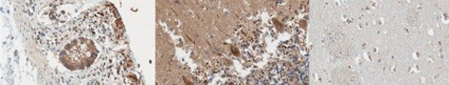
| Application【应用】 | Research Category Signaling Immunohistochemistry Analysis: A 1:250-1,000 dilution from a representative lot detected cytochrome P450 26A1 in human cerebellum, cerebral cortex, and large intestine tissue sections. Western Blotting Analysis: 4 µg/mL from a representative lot detected cytochrome P450 26A1 in 10 µg of human liver microsome lysate. Immunofluorescence Analysis: A representative lot immunostained dentate gyrus MAP2-positive neurons, but not GFAP-positive glia, nor CA1 hippocampal astrocytes and microglia by fluorescent immunohistochemistry staining of formalin-fixed, paraffin-embedded human hippocampus and cerebellum tissue sections (Stoney, P.N., et al. (2015). Brain Struct. Funct. In press). Immunohistochemistry Analysis: Representative lots detected differential cytochrome P450 26A1 immunoreactivity among formalin-fixed, paraffin-embedded tissue sections from normal colon, colon cancer, and lymph node metastasis (Brown, G.T., et al. (2014). PLoS One. 9(3):e90776; Kumarakulasingham, M., et al. (2005). Clin. Cancer Res. 11(10):3758-3765). Immunohistochemistry Analysis: A representative lot detected a significantly greater cytochrome P450 26A1 immunoreactivity among ormalin-fixed, paraffin-embedded ovarian cancer tissue sections when compared with normal ovary tissue sections (Downie, D., et al. (2005). Clin. Cancer Res.11(20):7369-7375). Western Blotting Analysis: A representative lot detected the ~54 kDa cytochrome P450 26A1 target band in human hippocampus tissue homogenate (Stoney, P.N., et al. (2015). Brain Struct. Funct. In press). Western Blotting Analysis: A representative lot detected the overexpression of cytochrome P450 26A1 exogenously transfected into human embryonic kidney cells (Brown, G.T., et al. (2014). PLoS One. 9(3):e90776). Anti-Cytochrome P450 26A1, clone F27P6A1, Cat. No. MABS1319, is a highly specific mouse monoclonal antibody that targets Cytochrome P450 26A1 and has been tested in Immunofluorescence, Immunohistochemistry (Paraffin), and Western Blotting. |
| Quality【质量】 | Evaluated by Western Blotting in human hippocampus tissue lysate. Western Blotting Analysis: 2 µg/mL of this antibody detected cytochrome P450 26A1 in 10 µg of human hippocampus tissue lysate. |
| Physical form【外形】 | Protein G purified. Format: Purified Purified mouse IgG2bκ in buffer containing 0.1 M Tris-Glycine (pH 7.4), 150 mM NaCl with 0.05% sodium azide. |
| Other Notes【其他说明】 | Concentration: Please refer to lot specific datasheet. |
产品性质
| Quality Level【质量水平】 | 100 |
| biological source【生物来源】 | mouse |
| antibody form【抗体形式】 | purified immunoglobulin |
| antibody product type | primary antibodies |
| clone【克隆】 | F27P6A1, monoclonal |
| species reactivity | human |
| technique(s) | immunofluorescence: suitable immunohistochemistry: suitable (paraffin) western blot: suitable |
| isotype【同位素/亚型】 | IgG2bκ |
| NCBI accession no.【NCBI登记号】 | NP_000774 |
| UniProt accession no.【UniProt登记号】 | O43174 |
| shipped in【运输】 | ambient |
产品说明
| Target description【目标描述】 | ~53/48 kDa observed. 56.20/48.56 kDa (isoform 1/2) calculated. Uncharacterized bands may be observed in some lysate(s). |
| Storage and Stability【储存及稳定性】 | Stable for 1 year at 2-8°C from date of receipt. |
| Disclaimer【免责声明】 | Unless otherwise stated in our catalog or other company documentation accompanying the product(s), our products are intended for research use only and are not to be used for any other purpose, which includes but is not limited to, unauthorized commercial uses, in vitro diagnostic uses, ex vivo or in vivo therapeutic uses or any type of consumption or application to humans or animals. |
安全信息
| Storage Class Code【储存分类代码】 | 12 - Non Combustible Liquids |
| WGK | WGK 1 |