


您的位置:首页 > 产品中心 > Anti-LBPA Antibody, clone 6C4
Anti-LBPA Antibody, clone 6C4
产品别名
Anti-LBPA Antibody, clone 6C4
LBPA
基本信息
| eCl@ss | 32160702 |
| NACRES | NA.41 |
| General description【一般描述】 | Lyso bis-phosphatidic acid (LBPA, Lysobisphosphatidic acid, L(bis)PA) is a lipid that is exclusively located in late endosomes and is used as a late endosome marker. One of the characteristic features of late endosomes is a complex system of internal membranes within the lumen that contains large amounts of the unique, poorly degradable LBPA, and thus forms a specialized membrane domain within endosomes. Late endosomes function not only as a major protein-sorting compartment, but also an obligatory station for LDL and other endocytosed ligands destined for degradation. Research shows that the LBPA-rich membranes within late endosomes regulate cholesterol transport, presumably by acting as a collection and distribution device. The genetic disease Niemann–Pick type C (NPC) is characterized by both lysosomal and endosomal storage disorders. In skin fibroblasts from NPC patients, cholesterol accumulation is seen within vesicles containing late endosome markers LBPA and Rab7. |
| Specificity【特异性】 | Target molecule is not species-specific. Clone 6C4 specifically detects LBPA, but not PC, PE, SM, PS, CL, PI, Sul, Chol, Cer, or CE purified from baby hamster kidney (BHK) cell lipid extract (Kobayahsi, T., et al. (1998). Nature. 392(6672):193-197). |
| Immunogen【免疫原】 | Endosomal membranes from baby hamster kidney fibroblasts (BHK) corresponding to Hamster LBPA. |
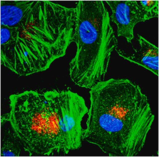
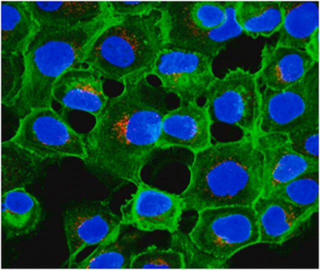
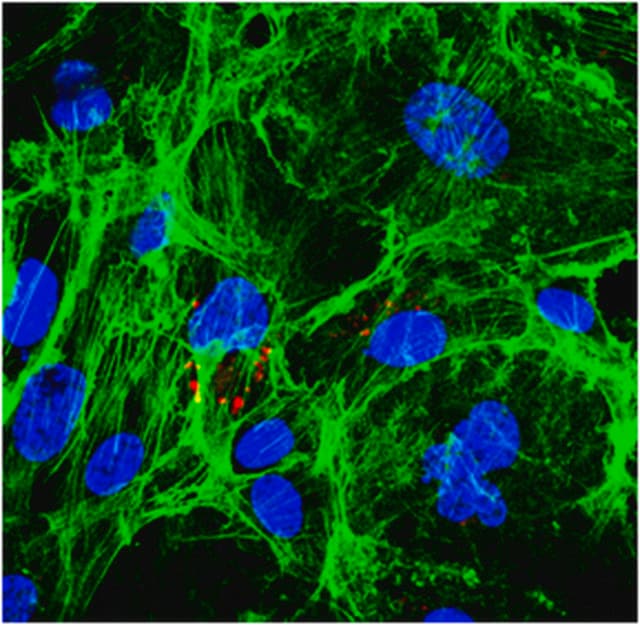
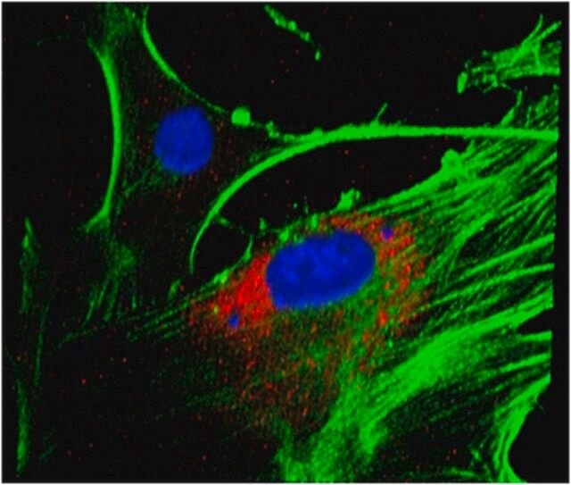
| Application【应用】 | Research Category Cell Structure Immunocytochemistry Analysis: 4.0 µg/mL from a representative lot detected LBPA in A431, HUVEC and NIH/3T3 cells. Immunocytochemistry Analysis: A representative lot detected late endosome LBPA immunoreactivity by fluorescent immunocytochemistry using MLN64-overexpressing MCF7 cells (Alpy, F., et al. (2001). J Biol Chem. 276(6):4261-4269). Immunocytochemistry Analysis: A representative lot detected late endosome LBPA immunoreactivity in skin fibroblasts from healthy donors, as well as Niemann–Pick type C (NPC) and Tay-Sachs disease patients by fluorescent immunocytochemistry (Kobayahsi, T., et al. (1999). Nat Cell Biol. 1(2):113-118). Immunocytochemistry Analysis: A representative lot detected late endosome LBPA immunoreactivity in baby hamster kidney (BHK) cells by fluorescent immunocytochemistry (Kobayahsi, T., et al. (1998). Nature. 392(6672):193-197). Electron Microscopy Analysis: A representative lot detected similar cellular LBPA distribution in the skin fibroblasts from autosomal recessive Niemann–Pick type C disease (NPC) patients as seen in baby hamster kidney (BHK) cells (Kobayahsi, T., et al. (1999). Nat Cell Biol. 1(2):113-118). Electron Microscopy Analysis: A representative lot detected LBPA immunoreactivity specifically associated with the late endosomes in baby hamster kidney (BHK) cells, but not the lgp120-positive limiting membranes of the same endosomes (Kobayahsi, T., et al. (1998). Nature. 392(6672):193-197). ELISA Analysis: A representative lot specifically detected wells coated with total baby hamster kidney (BHK) cell lipid extract or purified LBPA, but not other lipids (PC, PE, SM, PS, CL, or PI) purified BHK lipid extract (Kobayahsi, T., et al. (1998). Nature. 392(6672):193-197) Dot Blot Analysis: A representative lot specifically detected purified LBPA spotted on a HPTLC plate, but not other lipids (PC, PE, SM, PS, CL, PI, Sul, Chol, Cer, CE) purified from baby hamster kidney (BHK) cell lipid extract (Kobayahsi, T., et al. (1998). Nature. 392(6672):193-197). This Anti-LBPA Antibody, clone 6C4 is validated for use in Immunocytochemistry, Electron Microscopy, ELISA and Dot Blot for the detection of LBPA. Research Sub Category Adhesion (CAMs) |
| Quality【质量】 | Evaluated by Immunocytochemistry in HeLa cells. Immunocytochemistry Analysis: 4.0 µg/mL of this antibody stained late endosomes in HeLa cells. |
| Physical form【外形】 | Purified mouse monoclonal IgG1κ antibody in buffer containing 0.1 M Tris-Glycine (pH 7.4), 150 mM NaCl with 0.05% sodium azide. Format: Purified Protein G Purified |
| Other Notes【其他说明】 | Concentration: Please refer to lot specific datasheet. |
产品性质
| Quality Level【质量水平】 | 100 |
| biological source【生物来源】 | mouse |
| antibody form【抗体形式】 | purified immunoglobulin |
| antibody product type | primary antibodies |
| clone【克隆】 | 6C4, monoclonal |
| species reactivity (predicted by homology) | all |
| technique(s) | ELISA: suitable dot blot: suitable electron microscopy: suitable immunocytochemistry: suitable |
| isotype【同位素/亚型】 | IgG1κ |
| shipped in【运输】 | wet ice |
产品说明
| Storage and Stability【储存及稳定性】 | Stable for 1 year at 2-8°C from date of receipt. |
| Disclaimer【免责声明】 | Unless otherwise stated in our catalog or other company documentation accompanying the product(s), our products are intended for research use only and are not to be used for any other purpose, which includes but is not limited to, unauthorized commercial uses, in vitro diagnostic uses, ex vivo or in vivo therapeutic uses or any type of consumption or application to humans or animals. |
安全信息
| Storage Class Code【储存分类代码】 | 12 - Non Combustible Liquids |
| WGK | WGK 1 |